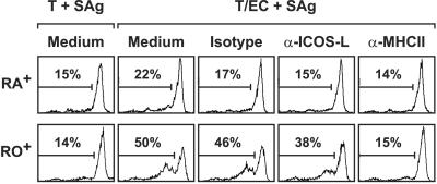
Figure 9.
Influence of EC-expressed ICOS-L on T cell proliferation. CD4+CD45RA+ or CD4+CD45RO+ T cells were labeled with carboxyfluorescein diacetate succinimidyl ester and cocultured with IFN-γ-stimulated HUVEC and SAg for 5 days in the absence or presence of an isotype control mAb (2A11), anti-ICOS-L mAb (HIL-131), or anti-MHC class II mAb (L243). T cells were analyzed for cell division by flow cytometry. Data are representative of two experiments. T, T cells.

