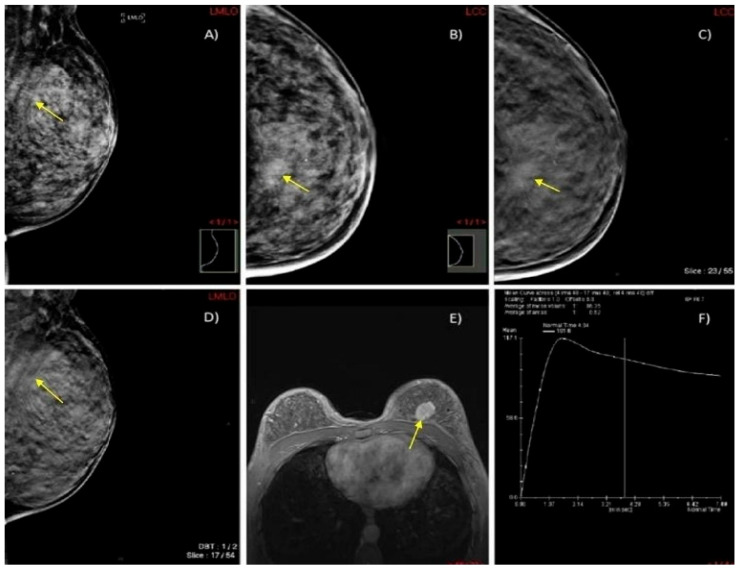
Figure 6.
A case of a 45-year-old woman with extremely dense breasts (ACR category D) and HPE-confirmed invasive ductal cancer in the left breast. (A,B) DM in MLO and CC projections shows an architectural distortion in the upper inner quadrant of the left breast (yellow arrow). (C,D) DBT in MLO and CC projections illustrates a spiculate mass in the upper inner quadrant of the left breast (yellow arrow). (E) Axial 3D T1-weighted FLASH fat-suppressed (FS) MR image demonstrates an extensive post-contrast hyperintense lesion in the prepectoral space of the left breast (yellow arrow). (F) MRI dynamic contrast enhancement curve type III (“wash out” curve).

