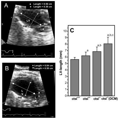
Figure 4.
In vivo assessment of left ventricular dimensions by echocardiography. (A) Two-dimensional echocardiographic picture of a normal-sized left ventricle in a 12-month-old wild-type mouse. Ao, aortic valve; LV, left ventricle; LA, left atrium; IVS, interventricular septum; PW, posterior wall. (B) Enlarged left ventricle of a 12-month-old CTSL-deficient mouse. (C) Genotype-dependent increase in the length of left ventricle. ctsl+/+, wild-type mice; ctsl+/−, heterozygous mice; ctsl−/−, CTSL-deficient mice; ctsl−/− DCM, CTSL-deficient mice with manifest DCM. (a) P < 0.05 compared with ctsl+/+, (b) P < 0.05 compared with ctsl+/−, (c) P < 0.05 compared with ctsl−/−.

