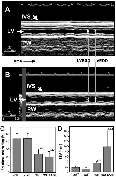
Figure 5.
Determination of heart contraction. (A) M-mode echocardiography showing normal contraction of the interventricular septum and the posteriolateral wall in the heart of a wild-type mouse. LV, left ventricle; IVS, interventricular septum; PW, posterior wall; LVEDD, left ventricular end-diastolic diameter; LVESD, left ventricular end-systolic diameter. (B) Reduced contraction of interventricular septum and posteriolateral wall in a CTSL-deficient mouse. (C) Fractional shortening as a measure of heart contraction and (D) left ventricular end-systolic volume in wild-type mice (ctsl+/+), heterozygous mice (ctsl+/−), CTSL-deficient mice (ctsl−/−), and CTSL-deficient mice with manifest DCM (ctsl−/− DCM). (a) P < 0.05 compared with ctsl+/+, (b) P < 0.05 compared with ctsl+/−, (c) P < 0.05 compared with ctsl−/−.

