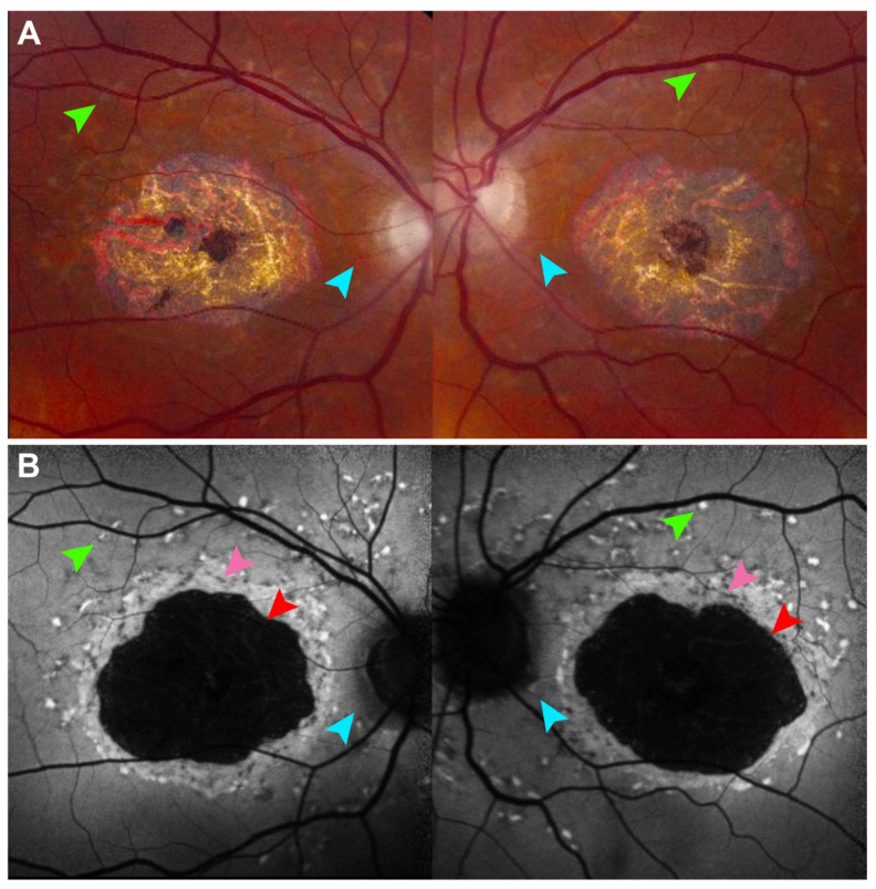
Figure 2.
Fundus autofluorescence in Stargardt disease detects features hard to see on color fundus photography. (A) 30° field color fundus photography of a 77-year-old patient with Stargardt disease demonstrates central chorioretinal atrophy and faint gray flecks (green arrows) around central atrophy and along the superior arcades. (B) 30° field fundus autofluorescence better demonstrates hyperautofluorescent flecks and peripapillary sparing around the optic nerve which is nearly imperceptible on fundus photography (cyan arrows). The well-demarcated definitely decreased autofluorescence (DDAF) (red arrow) has been used in multiple clinical trials to assess treatment efficacy in Stargardt disease. The questionably decreased autofluorescence (QDAF) (pink arrow) refers to areas with intermediate autofluorescence signal loss and represents a transitional stage between healthy retina and advanced atrophy. QDAF regions are often poorly demarcated and progress to DDAF over time.

