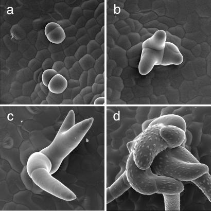
Figure 2.
Analysis of pGL2∷CYCD3;1 trichome development. (a–d) Scanning electron micrographs of pGL2∷CYCD3;1 trichomes. (a) Very young dividing trichomes giving rise to trichome clusters. (b and c) Further cell divisions take place as trichomes grow out and elongate. (d) Mature multicellular trichome comprising many cells. Note that the individual cells form papillae.

