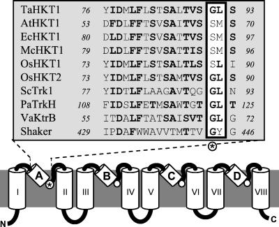
Figure 2.
Structural model of HKT proteins. Model of HKT/Trk/KtrB transporters as in refs. 13 and 18, based on the known structure of K+ channels (5). Predicted P-loops are labeled A–D, transmembrane domains are labeled I–VIII. The alignment shows P-loop A of various plant HKTs compared with Trk1 from S. cerevisiae (M21328), TrkH from Pseudomonas aeruginosa (AAG06598), KtrB from Vibrio alginolyticus (BAA32063), and to the P-loop of the Drosophila Shaker channel (S00479). The residue corresponding to the first glycine of the K+ channel GYG motif is marked with an asterisk.

