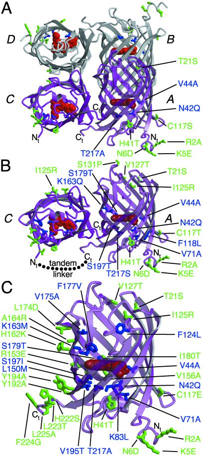
Figure 1.
Graphical representation of the tetramer, dimer, and monomer of DsRed based on the x-ray crystal structure of DsRed (21). Residues 1–5 were not observed in the crystal structure (Protein Data Bank identification 1G7K) but have been arbitrarily appended for the sake of representation. The DsRed chromophore is represented in red, and the four chains of the dimer are labeled following the convention of Yarbrough et al. (21). (A) The tetramer of DsRed with all residues mutated in T1 indicated in green for external residues and blue for those internal to the β-barrel. (B) The AC dimer of DsRed with all mutations present in dimer2 represented as in A and the intersubunit linker present in tdimer2(12) shown as a dotted line. (C) The monomer of DsRed with all mutations present in mRFP1 represented as in A. This figure was produced with molscript (27).

