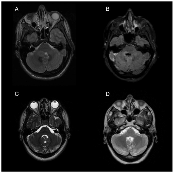
Figure 1.
Pre- and post-operative magnetic resonance imaging (MRI) of two patients with posterior fossa neoplasms. (A) Pre-operative T2 sequence of Patient 1, showing a 1.9 × 1.5 cm ovoid mass in the right cerebellar hemisphere extending into the fourth ventricle. (B) Post-operative T2 FLAIR image showing postsurgical changes with pontine and medullary hyperintensity. (C) Pre-operative T2 from Patient 2 showing an iso-intense mass partially obstructing the fourth ventricle and extending from the cerebellar vermis. (D) Post-operative T2 showing postsurgical volume loss of the inferior vermis.

