Figure 5.
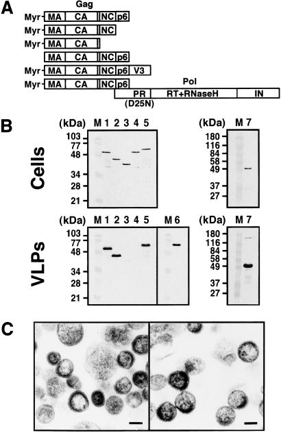
VLP formation of various Gag constructs. (A) Schematic representation of the Gags constructed. (B) Intracellular Gag expression and purified VLPs. Yeast cells (0.5 OD) expressing each Gag construct were subjected to SDS/PAGE followed by Western blotting (Upper). After removal of the cell wall, the spheroplasts were cultured for 2 h. VLPs were purified from the culture medium by sucrose gradient centrifugation and detected by Western blotting (Lower). Western blotting was carried out using anti-HIV-1 CA (for lanes 1–5 and 7) and V3 (for lane 6) Abs. Lanes: M, prestained molecular weight markers; 1, Myr-MA-CA-p2-NC-p6 (wild-type); 2, Myr-MA-CA-p2-NC; 3, Myr-MA-CA-p2; 4, nonmyristoylated MA-CA-p2-NC-p6; 5 and 6, Myr-MA-CA-p2-NC-p6-V3 fusion; and 7, gag-pol (containing the inactive form of protease). (C) Electron micrographs of VLPs. (Left) VLPs produced by expression of Myr-MA-CA-p2-NC. (Right) VLPs produced by expression of Myr-MA-CA-p2-NC-p6-V3 fusion. (Scale bars, 100 nm.)

