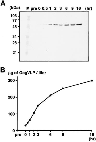
Figure 6.
Time courses of VLP production. After removal of the cell wall, yeast spheroplasts expressing the full-length Gag protein were cultured at 30°C. At each time point, VLPs were purified from the culture medium by sucrose gradient centrifugation. (A) VLP fractions detected by Western blotting using anti-HIV-1 CA Ab. Lanes: M, prestained molecular weight markers; pre, before removal of the cell wall; 0, just after removal of the cell wall; and 0.5–16, hours of cultivation following the removal. (B) VLP yields estimated by quantitative Western blotting and Coomassie brilliant blue staining. The VLP yield at each time point was estimated by comparison to standard dilutions prepared with purified soluble Gag protein (for Western blotting) or BSA (for Coomassie brilliant blue staining).

