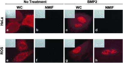
Figure 1.
Differential subnuclear organization of Smad5 in nonosseous and osseous cells. Human cervical carcinoma HeLa cells (a–d) or rat osteosarcoma ROS 17/2.8 cells (e–h) were grown on gelatin-coated coverslips and transfected with 0.5 μg of expression construct coding for Flag Smad5. After 24 h of transfection, cells were processed in situ for the WC and the NM-IF preparation and immunofluorescence microscopy. A mouse mAb against Flag tag (dilution: 1:1000) was used to detect Smad5.

