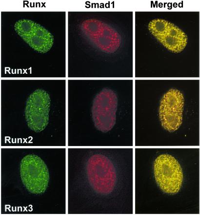
Figure 3.
Runx factors specify subnuclear localization of Smads. HeLa cells were transfected with 0.5 μg of expression construct for Flag Smad1 along with Runx1, Runx2, or Runx3 expression vectors. Cells were treated with BMP2, and Smad1 was examined for association with Runx factors in the nuclear matrix (NM-IF preparation) by in situ immunofluorescence 24 h after transfection. Smad1 was detected with mouse mAb against Flag tag. Runx proteins were detected with rabbit polyclonal Abs at a dilution of 1:200. Images were taken by a Zeiss Axioplan microscope coupled with a charge-coupled device (CCD) camera and were processed for deconvulation microscopy with METAMORPH bioimaging software.

