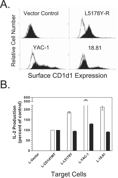
Figure 2.
Functional cell surface expression of CD1d1 on L cells transfected with cd1d1 cDNA from murine CD1+ hematopoietic tumor cells. (A) Murine L cell fibroblasts were transfected with the pcDNA3.1-neo vector alone or the vector containing the full-length cd1d1 cDNA generated from L5178Y-R, YAC-1, or 18.81 cells. The cells were stained with a PE-labeled anti-mouse CD1 mAb (filled histograms). A PE-conjugated rat IgG2b (open histograms) served as an isotype control. Analysis was by cytofluorography. The data are representative of two independent experiments. (B) L cells transfected with vector only or vector containing the WT cd1d1 cDNA (L-CD1d1WT) or that from L5178Y-R, YAC-1, or 18.81 cells were cocultured with the Vα14+ (canonical; DN32.D3; white bars) or Vα5+ (noncanonical; N37-1A12; black bars) NKT cell hybridomas for 24 h. Supernatants were harvested, and IL-2 production was measured by ELISA. The data shown are the mean of triplicate cultures ± SD.

