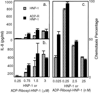
Figure 4.
Effects of ADP-ribosyl-HNP-1 and HNP-1 on IL-8 release by A549 cells and on T cell chemotaxis. Cells were incubated for (a) 12 or (b) 24 h with the indicated concentration of HNP-1 or ADP-ribosyl-HNP-1 before analysis of medium. IL-8 concentration in medium from cells incubated without defensins has been subtracted (at 12 h, 36.4 ± 6 pg/ml; at 24 h, 85 ± 34 pg/ml). Data are means ± 1/2 range of values from two experiments, each performed in triplicate. *, P < 0.05 for difference between IL-8 release stimulated by HNP-1 and ADP-ribosyl-HNP-1. CD3+ cells were incubated with the indicated concentrations of HNP-1 or ADP-ribosyl HNP-1 (c). We used MIP-1β (5 ng/ml) in migration medium, or migration medium alone, as positive and negative controls, respectively. Chemotaxis percentage was calculated as: (number of cells migrated to the lower chamber in the experimental conditions − number of cells migrated in the negative control)/(number of cells migrated in the positive control − number of cells migrated in negative control) × 100. Data are means ± SEM values from four experiments, each performed in duplicate.

