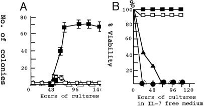
Figure 4.
Colony formation of BM cells by transfection of the expression vector carrying Stat5a cDNA. (A) Kinetics of the colony formation of BM cells transfected with Stat5a cDNA. The “numbers of colony” represent colonies formed by 2.8 × l06 transfected BM cells. ■, Colonies of BM cells transfected with 4 μg of the 1*6-Stat5a expression vector; □, colonies of SL/Kh BM cells transfected with 4 μg of the wt Stat5a expression vector. The dose of cDNA was between 2 μg and 8 μg for 2 × l07 BM cells. ▵, Colonies from SL/Kh BM cells transfected with 4 μg of the empty vector. (B) The survival of the colony cells in the IL-7-free medium. The colony cells harvested 96 h after transfection of 1*6-Stat5a were incubated in a medium with (■) or without (□) IL-7. Similar cells harvested 48 h after transfections were incubated in the medium with (▴) or without (▵) IL-7. These cells died quickly of apoptosis. The viability of the BM cells transfected with wt Stat5a was as low as 10% when harvested 48 or 72 h after transfection and no cells survived after transfer to IL-7-free medium (●).

