Figure 4.
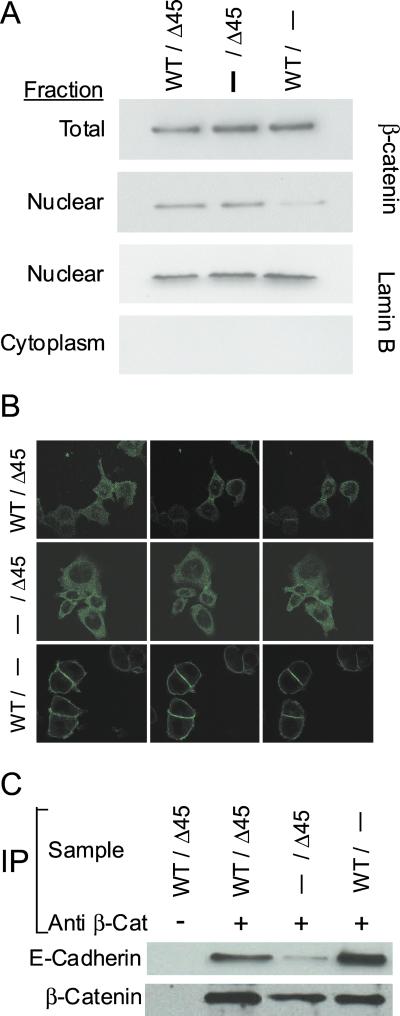
Mutant β-catenin displays abnormal subcellular localization and decreased binding to E-cadherin. (A) Cells with the indicated genotypes were fractionated and analyzed by Western blotting as indicated. The total amount of β-catenin is similar in parental, WT (−/Δ45), and mutant (WT/−) KO clones. WT β-catenin is largely excluded from the nuclear fraction, whereas mutant β-catenin is not. Western blot analysis of the nuclear protein lamin B controlled for the quality of the fractionation. (B) Parental (WT/Δ45), WT (−/Δ45), and mutant (WT/−) CTNNB1 KO cells were stained with anti-β-catenin antibody and imaged by using a confocal microscope. Confocal images at three different levels are shown for each cell type. (C) Lysates from KO cells were immunoprecipitated with anti-β-catenin antibody as indicated. The immunoprecipitated complexes were then analyzed by Western blotting using anti-E-cadherin antibody or anti-β-catenin. Mutant β-catenin has decreased binding to E-cadherin (compare −/Δ45 with WT/− and WT/Δ45).

