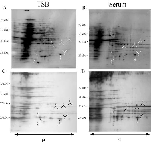
FIG. 1.
Protein expression profiles of cell wall fractions from S. epidermidis 0-47 grown in TSB (A and C) or 70% rabbit serum (B and D) were compared by 2D gel electrophoresis. Prior to fractionation of the cell wall-associated proteins from the cells grown in serum, the cells were washed with a buffer containing 0.5 M NaCl to remove serum proteins. The TSB cells were treated the same. Proteins were separated on pH 4 to 7 IPG strips followed by SDS-PAGE, transferred to nitrocellulose, and detected by fluorescent stain (A and B). Immunoreactive proteins were visualized with immune sera (C and D) from rabbits immunized with S. epidermidis 0-47. Spots or streaks expressed at different levels in the presence of serum are labeled a to h. Molecular mass markers are shown on the left.

