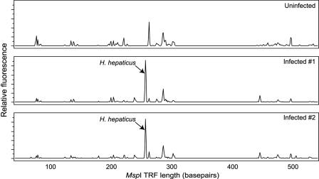
FIG. 1.
Representative T-RFLP traces comparing the mucosa-associated microbiota in the cecae of an uninfected C57BL/6 mouse and two mice infected with H. hepaticus 30 days previously. The relative fluorescence of each peak is plotted against the size of the peak. In the traces from the two infected animals, a TRF corresponding to an H. hepaticus-specific TRF is clearly seen and is the dominant TRF.

