FIG. 1.
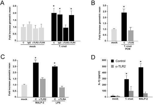
TLR activation triggers cytokine production and hypertrophy in isolated cardiomyocytes. (A) Cardiomyocytes incubated with medium alone (control [C]), control IgG (10 μg/μl) (IgG), anti-TLR2 IgG (10 μg/μl) (αTLR2), or anti-TLR4 IgG (20 μg/μl) (αTLR4) were mock-treated or infected with T. cruzi for 48 h. Hypertrophic cells were detected by flow cytometry, where a ≥2-fold change in the geometric mean of FSC indicates a significant increase in cell size for cardiomyocyte populations. FSC data from all treatments and controls were normalized to mock-treated cells incubated in medium alone (i.e., mock, C), for which the average FSC value was set at 1.0. Data are represented as the averages for five independent experiments carried out in duplicate ± the standard deviation. Asterisks denote a significant change from mock-treated controls (i.e., mock, C) at a P of <0.05 via Student's t test with Welch correction for multiple comparisons. (B and C) Cardiomyocytes were mock-treated or stimulated with PCM (B) or MALP-2 (10−7 U/ml) and LPS (5 ng/ml) (C) in the presence or absence of TLR-blocking antibodies. (D) ELISA analysis of IL-1β in medium harvested from mock-treated (mock) cardiomyocytes or cells exposed to T. cruzi MALP-2 trypomastigotes for 48 h in the absence (Control) or presence (αTLR2) of TLR2-blocking antibody. Data represents an average of three independent experiments carried out in triplicate ± the standard deviation. Asterisks denote a significant change from mock-treated cells (P < 0.05) via Student's t test with Welch correction for multiple comparisons.

