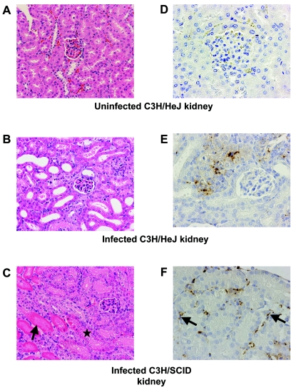
FIG. 2.
Mouse kidney tissue infected with Leptospira. A hematoxylin and eosin stain of a normal C3H/HeJ kidney (A), compared to an infected C3H/HeJ kidney (B) and an infected C3H/SCID kidney (C), is shown (original magnification, ×200). Infected kidneys show evidence of tubular injury. Frankly necrotic renal tubules are indicated by arrows compared to a renal tubule with early necrotic changes (asterisk). The immunohistochemistry of a negative-control kidney (D), compared to an infected C3H/HeJ kidney with granular debris (E) and infected C3H/SCID kidney with intact leptospires (arrows) (F), is shown (original magnification, ×200).

