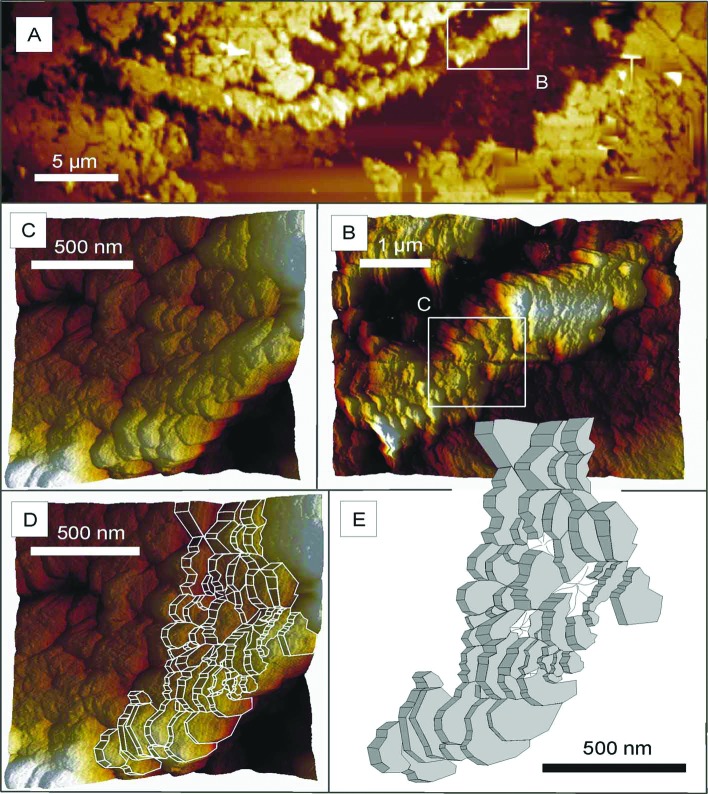
Figure 3.
Atomic force micrographs of a portion of the petrified Chichkan sphaeromorph. (A) The portion of the cell wall imaged in B–D and illustrated in E, a segment of the wall shown in the lowermost part of Fig. 2B. (B) That portion of the wall indicated by the white rectangle in A, shown here at higher magnification. (C) That portion of the wall indicated by the white rectangle in B, shown here at higher magnification. (D) That portion of the wall shown in C with a superimposed computer-generated outline of its component parts. (E) A computer-generated sketch of the platelet-like wall subunits.

