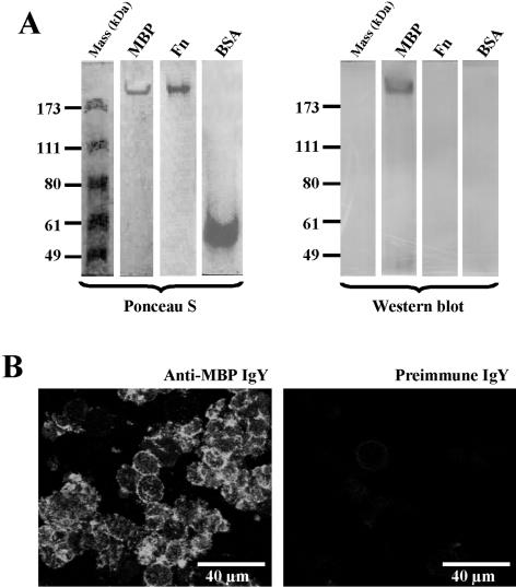
FIG. 2.
Western blot analysis and immunohistochemical localization of Acanthamoeba MBP. (A) Affinity-purified amoeba MBP was electrophoresed under native conditions. After electrophoresis, the proteins were transferred onto nitrocellulose membranes; the protein blots were stained with Ponceau S and then processed for immunostaining with anti-MBP IgY. The IgY antibody reacted with the MBP, but not with ferritin (Fn), BSA, or molecular mass markers. Also the MBP did not react with preimmune IgY (not shown). (B) Trophozoites (106 cells) were immunostained in cell suspension using anti-MBP IgY (left) or preimmune IgY (right) and FITC-labeled goat anti-chicken IgY. After being immunostained, the cells were smeared onto glass slides, fixed with 2.5% paraformaldehyde, and visualized under the confocal microscope.

