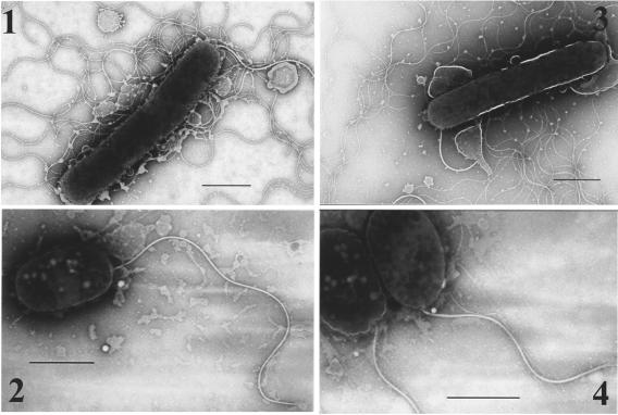
FIG. 5.
Transmission electron microscopy images of wild-type and mutant strains. Bacteria were grown on the LB plate supplemented with 3% NaCl at 37°C overnight and gently placed onto Formvar-coated copper grids and negatively stained using 2% potassium phosphotungstate. 1, wild-type strain; 2, vpaH deletion mutant strain; 3, vpaH complementation strain; 4, lafTU deletion mutant strain. Bars, 1 μm.

