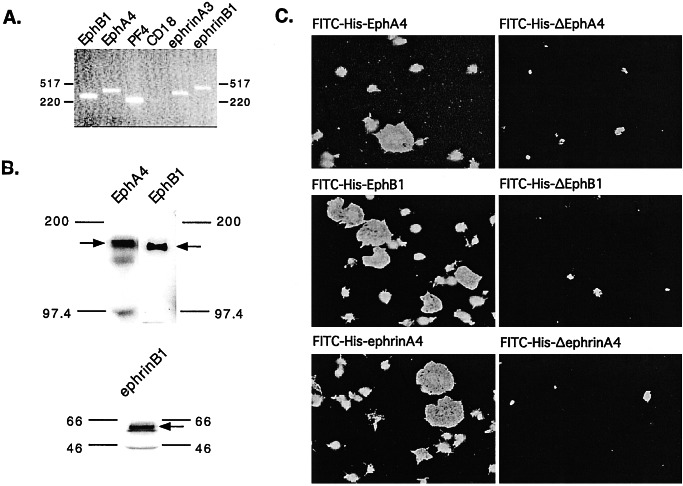
Figure 1.
Evidence for Eph kinases and ephrins on the surface of human platelets. (A) Reverse transcription and amplification (RT-PCR) of platelet RNA using primers specific for EphB1, EphA4, ephrinA3, and ephrinB1. Platelet factor 4 (PF4) is specific for platelets, whereas CD18 was used as a marker for leukocyte contamination. Sequencing of the amplified products confirmed their identities. (B) Immunoblots of total platelet lysates with antibodies that recognize EphA4, EphB1, and ephrinB1. (C) Platelets were incubated with 100 nM phorbol 12-myristate 13-acetate, allowed to adhere and spread on a fibrinogen-coated surface, and then stained with FITC-conjugated, His-6-tagged recombinant proteins corresponding to the exodomains of EphA4, EphB1, and ephrinA4. Control proteins lacked residues required for Eph/ephrin interactions are denoted with a “Δ.”

