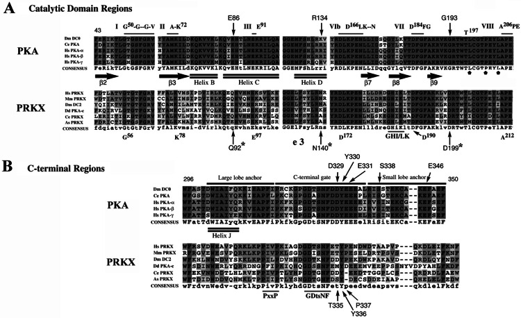
Figure 2.
Sequence alignment of PKA and PRKX family members. (A) Conserved residues of kinase domains I, II, III, VIb, VII, and VIII (horizontal lines) of the catalytic core domain (10), the PKA β-strands β2, β3, β7, β8, and β9 (filled arrows), the α-helices B, C, and D (double lines), and the PKA T197 autophosphorylation site are shown. The PRKX residues (G92, N140, and D199), which differ from PKA residues involved in RI subunit binding, are indicated by asterisks; PKA substrate P+1 recognition residues L198, P202, and L205 (domain VIII) are designated by small, filled pentagons. (B) C-terminal region alignments for PRKX and PKA families are shown with the large and small lobe anchor and C-terminal gate of PKA designated (33). Regions of conservation within the PRKX family shared with the PKA family are designated by the consensus sequences PxxP and GDtsNF.

