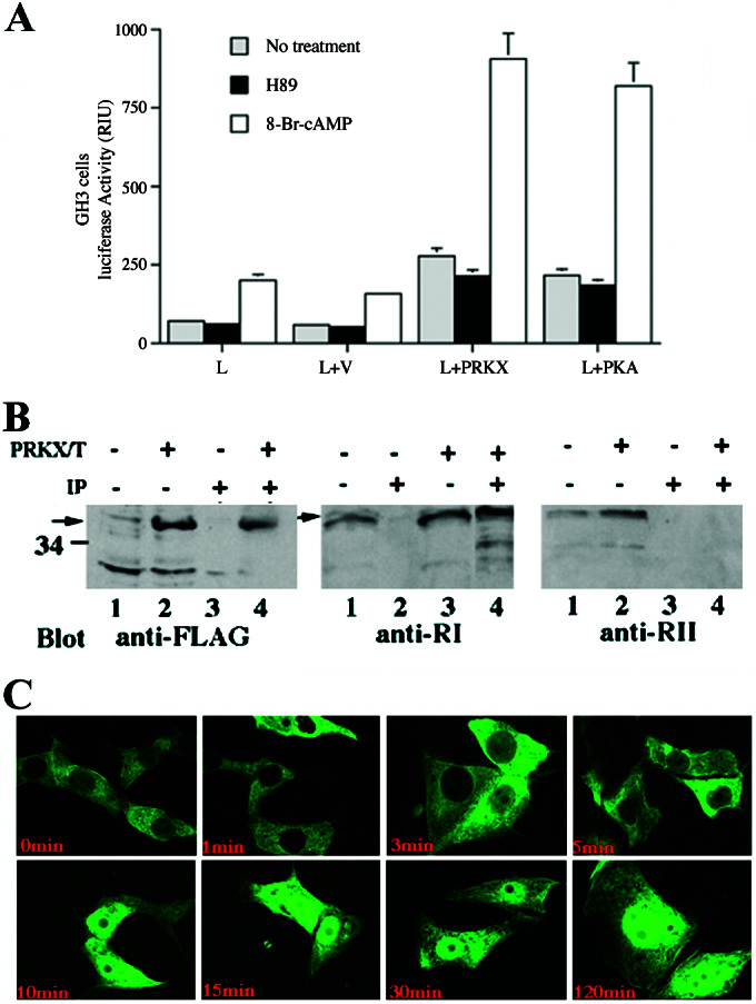
Figure 4.

cAMP stimulation of PRKX-dependent CRE promoter elements, detections of PRKX–RI holoenzyme complexes, and nuclear translocation of EGFP/PRKX fusion protein. (A) Five micrograms of PRKX expression construct cotransfected with 3 μg of pCRE-luciferase into JAR cells incubated in the presence or absence of 1 μM 8-Br-cAMP or 1 μM H89 followed by luciferase activity assay 24 h after transfection. The letters on the abscissa refer to constructs used for cell transfection (L, CRE-luciferase promoter-reporter; V, empty expression vector; PRKX, pFLAG/PRKX; PKA, pFC-PKA). All values are means ± SEM for two separate transfection experiments carried out in triplicate. (B) PKRX–RI complexes were detected by immunoprecipitation experiments of PRKX-transfected cells. PRKX/T (+, −) indicates cell transfection with pFLAG/PRKX. IP (+, −) indicates immunoprecipitation with anti-FLAG antibody beads. Lanes 1 and 2 (Left and Right) and lanes 1 and 3 (Middle) represent immunoblots performed on whole-cell lysates. Lanes 3 and 4 (Left and Right) and lanes 2 and 4 (Middle) are immunoblots performed on anti-FLAG immunoprecipitated proteins. (C) cAMP-dependent PRKX nuclear translocation demonstrated in FIB4 cells transfected with pEGFP/PRKX and treated with 100 μM 8-Br-cAMP for 0, 1, 3, 5, 10, 15, 30, and 120 min.
