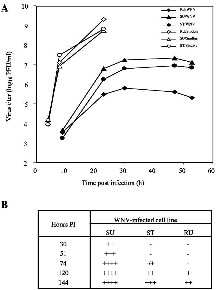
Figure 4.
Effect of the low level expression of the resistant Oas1b protein in C3H/He cells on the growth of a flavivirus, WNV, and an alpha togavirus, Sindbis. (A) Virus growth curves. Cells were infected with either WNV or Sindbis virus at a multiplicity of infection of 0.5. Samples of culture fluid were taken at the indicated times and titered by plaque assay on BHK cells. RU, untransfected resistant C3H.PRI-Flvr cells; SU, untransfected susceptible C3H/He cells; ST, susceptible C3H/He cells stably transfected with Oas1b cDNA from resistant C3H.PRI-Flvr cells. (B) Time course of the development of CPE after infection of SU, RU, and ST cells with WNV. −, no obvious CPE; +, rounding or detachment of ≈25% of the cells in the monolayer; ++, rounding or detachment of ≈50% of the cells in the monolayer; +++, rounding or detachment of ≈75% of the cells in the monolayer; ++++, complete destruction of the monolayer. PI, postinfection.

