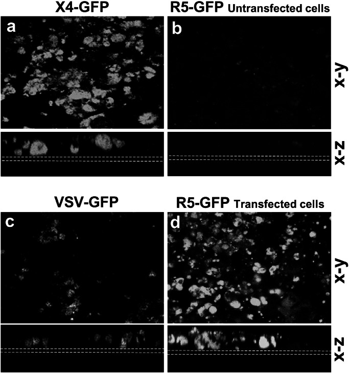
Figure 2.
Chemokine receptors specify HIV-1 strain specificity of infection. Confocal microscopy (x–y and x–z axes) of untransfected Caco-2 monolayers infected at 9 days with HxB2 X4 (a), ADA R5 HIV-1 (b), or VSV as a positive control (c). On integration in the host cell, the virus produces the GFP protein. On day 8 postinfection the filters were examined for GFP expression. Caco-2 infected with X4 (a) and with VSV (c) were GFP-positive, whereas monolayers infected with R5 remained negative (b). In contrast CCR5-transfected Caco-2 monolayers when exposed to R5 HIV-1 were positive for GFP expression (d), indicating that R5 successfully infected the CCR5-transfected Caco-2 cells. Dotted white lines indicate the position of the filters. (Bar = 20 μm.)

