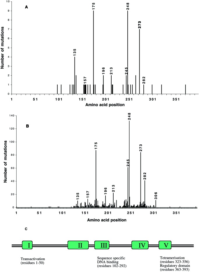
Figure 2.
Spectrum of p53 mutations in colorectal cancer. (A) p53 mutations in Dundee patients with colorectal cancer and (B) p53 mutations in all white colorectal solid tumors in the IARC p53 mutation database (18). The amino acid positions of the most frequently mutated hotspot codons are highlighted. (C) The position of each of the hotspot codons relative to the conserved regions and functional domains of p53 is illustrated.

