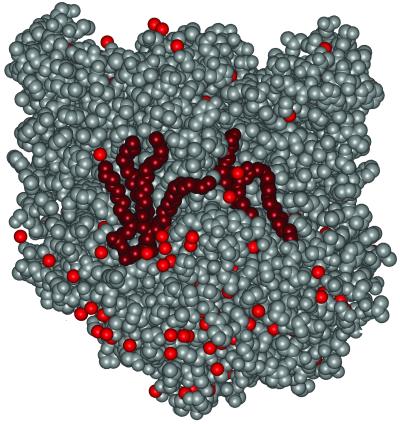
Fig 4.
Surface representation of the RC structure showing the glycolipid and cardiolipid (dark red), the protein (gray), and bound water molecules (light red). Both of the lipids are tightly bound, with the glycolipid fitting between the M and H subunits whereas the cardiolipin is more exposed but held by electrostatic interactions involving the two phosphates and nearby charged residues. The two lipids are positioned with a separation of about 30 Å between the phosphate and saccharide moieties.

