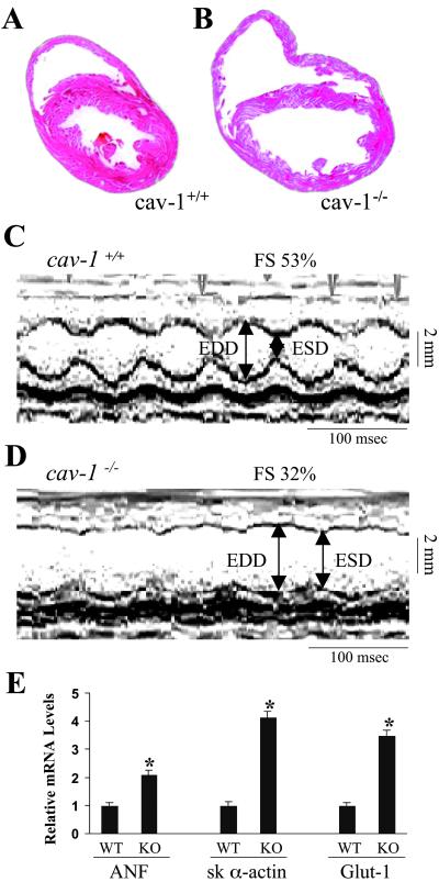
Fig 3.
Pathological analysis of the cav-1 knockout hearts. (A and B) Histological sections of hearts from 5-month-old littermates. Hearts were fixed in paraformaldehyde and stained with hematoxylin and eosin. Data are representative of three independent experiments with nearly identical results. (C and D) Transthoracic M-mode echocardiographic tracings in a wild-type mouse (C) and cav-1 knockout mouse (D). LV dimensions are indicated by the double-sided arrows. EDD, end diastolic dimension; ESD, end systolic dimension. Cav-1 knockout mice have chamber dilation with reduced wall motion, indicating depressed cardiac function and increased wall stress. (E) Quantitative analysis of the expression levels of ANF, skeletal α-actin and glut-1 genes in the heart by real time RT-PCR. The data were expressed as mean ± SEM. *, P < 0.05. ANF, proatrial natriuretic factor; sk α-actin, skeletal α-actin; Glut-1, glucose transporter-1.

