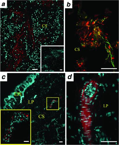
Fig 5.
MC4R expression in nerve fibers, nerve endings, and sensory corpuscles of the human glans penis. In situ hybridization was carried out by using riboprobes for human (a–c) or rat (d) MC4R. (a) MC4R mRNA in the human penis corpus spongiosum (CS) is seen in nerve fibers (shown in red); nuclei are seen in blue. Sense control probe produced no specific signal (Inset). (b) MC4R colocalized with the nerve fiber marker PGP9.5 in a CS mechanoreceptor. MC4R signal is shown in green, PGP9.5 immunoreactivity in red, and colocalization in yellow. (c) MC4R in situ hybridization signal in an encapsulated sensory corpuscle of the CS of the human glans penis. MC4R is shown in red, nuclei in blue. Layers of the human glans penis are labeled as epithelium (Epi), lamina propria (LP), and CS. High magnification view (Inset) suggests that this is a Pacinian-lamellated corpuscle. (d) MC4R localization in a Meissner-encapsulated corpuscle found in the LP of the rat glans penis. MC4R signal is shown in red, nuclei in blue. (Bars = 20 μm.)

