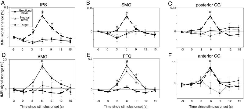
Fig 2.
Mean signal change (±SEM) in posterior brain regions. (Upper) Dorsal regions are presented in the (A) intraparietal sulcus (IPS), (B) supramarginal gyrus (SMG), and (C) posterior cingulate (CG). (Lower) Ventral regions are presented in the (D) amygdala (AMG), (E) fusiform gyrus (FFG), and (F) anterior CG (from 18.75 to 7.5 mm rostral to the AC). Data from the right and left hemispheres are collapsed. Note change in vertical scale across regions. Asterisks indicate time points where (A–C) targets evoked more activation than distracters, (D) emotional distracters evoked more activation than neutral distracters or targets, (E) distracters evoked more activation than targets, and (F) targets and emotional distracters evoked more activation then neutral distracters. The pound sign in E indicates where emotional distracters elicited stronger responses than neutral distracters, which in turn elicited stronger responses than targets.

