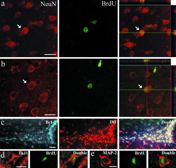
Fig 1.
Colocalization of neurogenesis markers in the rostral portion of the temporal lobe of squirrel monkeys that were killed 21 and 28 days after the last BrdUrd injection. Examples of a cell in the amygdala (a) and piriform cortex (b) that have incorporated BrdUrd (green) and also express NeuN (red). The right side shows reconstructed orthogonal images of the same NeuN+/BrdUrd+ cells, which are viewed from the sides in both x–z (top) and y–z (right) planes. (c) Cells in TS are colabeled for DiI (red) and for Bcl-2 (green). (d) TuJ1+/BrdUrd+ cell in TS and (e) MAP-2+/BrdUrd+ cell in amygdala. The various neuronal markers are stained in red, whereas BrdUrd is in green. [Bars = 25 μm (a and b), 100 μm (c), 15 μm (d and e).]

