

Abstract
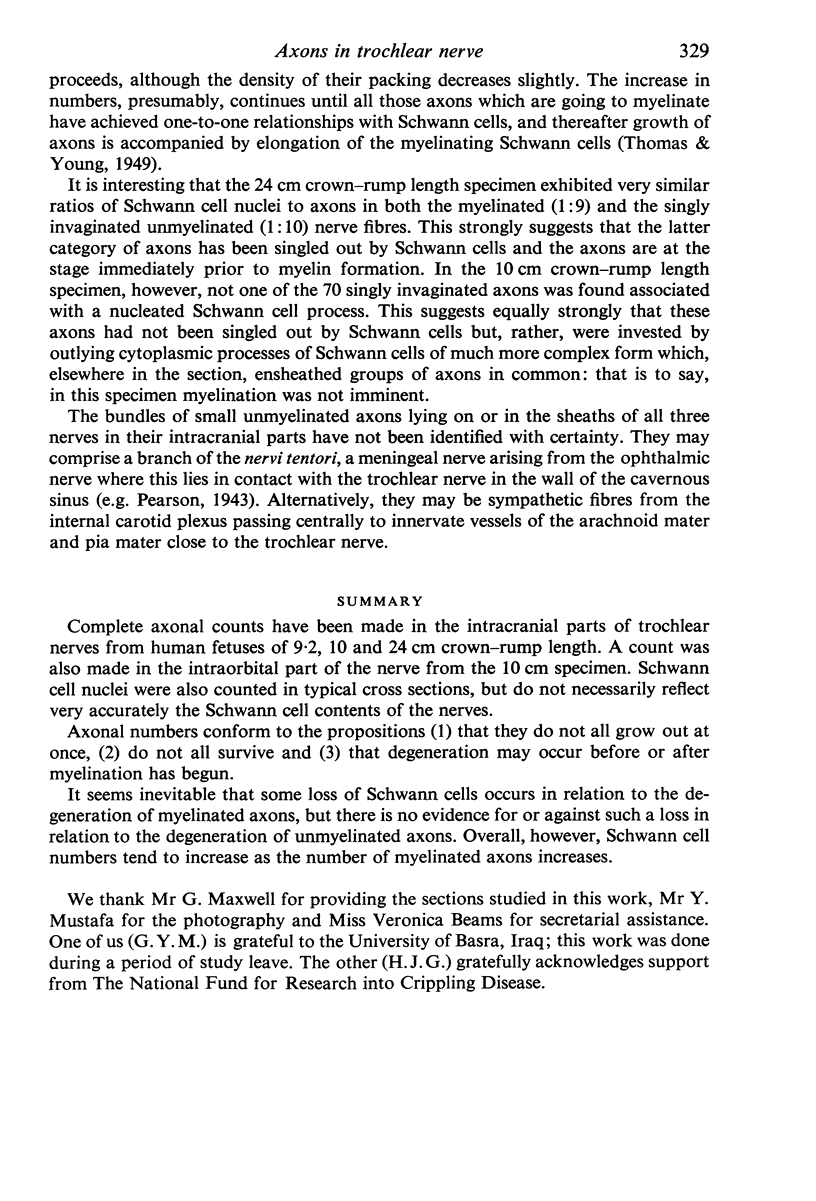
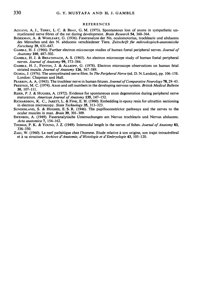
Complete axonal counts have been made in the intracranial parts of trochlear nerves from human fetuses of 9.2, 10 and 24 cm crown-rump length. A count was also made in the intraorbital part of the nerve from the 10 cm specimen. Schwann cell nuclei were also counted in typical cross sections, but do not necessarily reflect very accurately the schwann cell contents of the nerves. Axonal numbers conform to the propositions (1) that they do not all grow out at once, (2) do not all survive and (3) that degeneration may occur before or after myelination has begun. It seems inevitable that some loss of Schwann cells occurs in relation to the degeneration of myelinated axons, but there is no evidence for or against such a loss in relation to the degeneration of unmyelinated axons. Overall, however, Schwann cell numbers tend to increase as the number of myelinated axons increases.
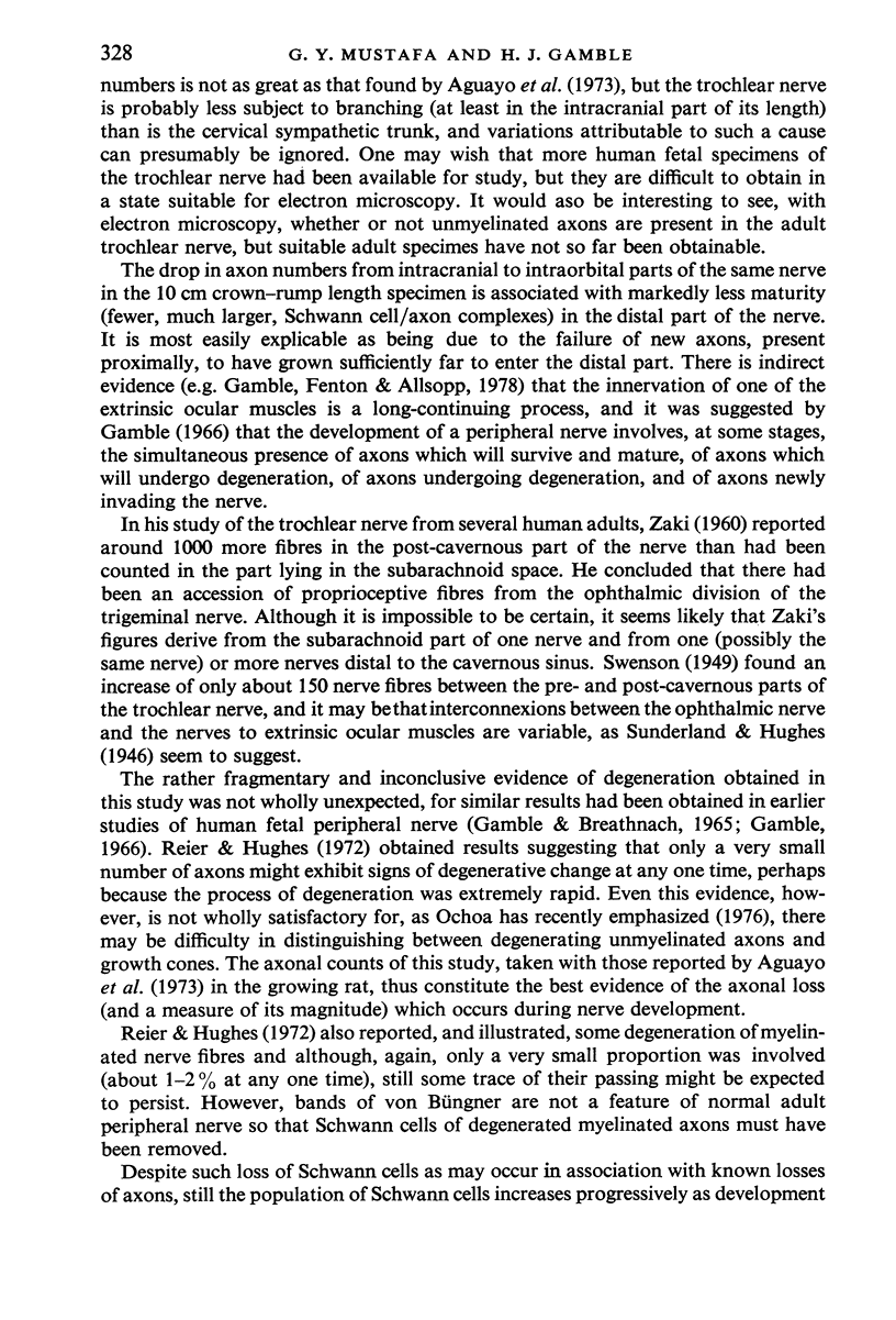
Full text
PDF


Images in this article
Selected References
These references are in PubMed. This may not be the complete list of references from this article.
- Aguayo A. J., Terry L. C., Bray G. M. Spontaneous loss of axons in sympathetic unmyelinated nerve fibers of the rat during development. Brain Res. 1973 May 17;54:360–364. doi: 10.1016/0006-8993(73)90061-9. [DOI] [PubMed] [Google Scholar]
- Gamble H. J., Breathnach A. S. An electron-microscope study of human foetal peripheral nerves. J Anat. 1965 Jul;99(Pt 3):573–584. [PMC free article] [PubMed] [Google Scholar]
- Gamble H. J., Fenton J., Allsopp G. Electron microscope observations on human fetal striated muscle. J Anat. 1978 Aug;126(Pt 3):567–589. [PMC free article] [PubMed] [Google Scholar]
- Gamble H. J. Further electron microscope studies of human foetal peripheral nerves. J Anat. 1966 Jul;100(Pt 3):487–502. [PMC free article] [PubMed] [Google Scholar]
- Prestige M. C. Axon and cell numbers in the developing nervous system. Br Med Bull. 1974 May;30(2):107–111. doi: 10.1093/oxfordjournals.bmb.a071178. [DOI] [PubMed] [Google Scholar]
- RICHARDSON K. C., JARETT L., FINKE E. H. Embedding in epoxy resins for ultrathin sectioning in electron microscopy. Stain Technol. 1960 Nov;35:313–323. doi: 10.3109/10520296009114754. [DOI] [PubMed] [Google Scholar]
- Reier P. J., Hughes A. Evidence for spontaneous axon degeneration during peripheral nerve maturation. Am J Anat. 1972 Sep;135(1):147–152. doi: 10.1002/aja.1001350113. [DOI] [PubMed] [Google Scholar]
- THOMAS P. K., YOUNG J. Z. Internode lengths in the nerves of fishes. J Anat. 1949 Oct;83(4):336-50, pl. [PMC free article] [PubMed] [Google Scholar]