Abstract
The epididymal region of the guinea-fowl was studied in sexually mature birds. The structure of the epididymal region was generally similar to that already described for the domestic fowl, turkey and Japanese quail. Well formed, intratesticular tubuli recti was seen connecting the seminiferous tubules with the rete testis. The latter consists of both intracapsular and extracapsular portions. Six main cell types were recognised in the region: the rete testis was lined by squamous cells, the proximal efferent ductules by ciliated and non-ciliated Type I cells, the distal efferent ductule by ciliated and non-ciliated Type II cells, and the connecting ductules, ductus epididymidis and ductus deferens were lined by non-ciliated Type III and basal cells. The cell classification adopted in this study is discussed.
Full text
PDF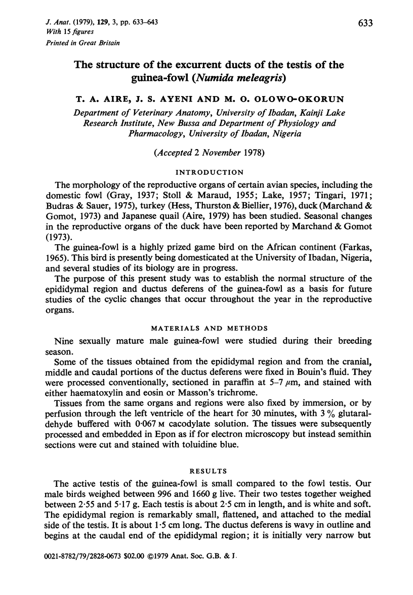
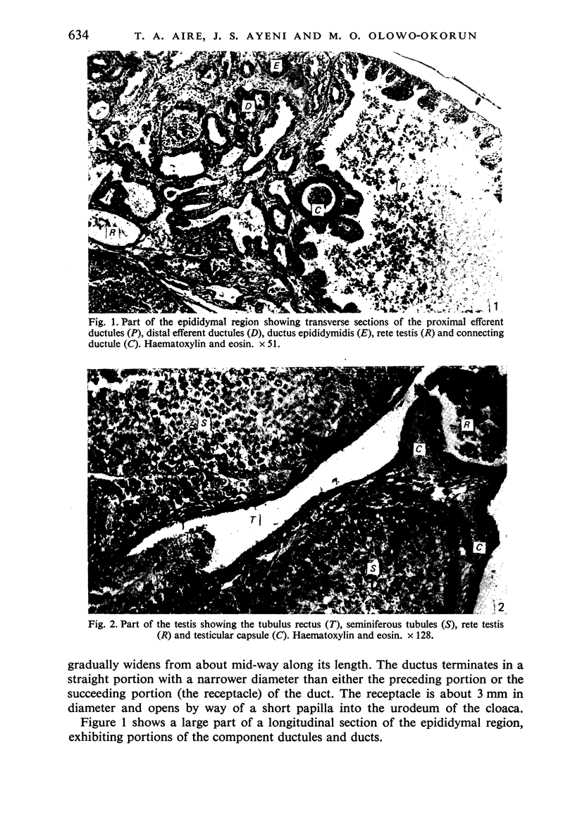
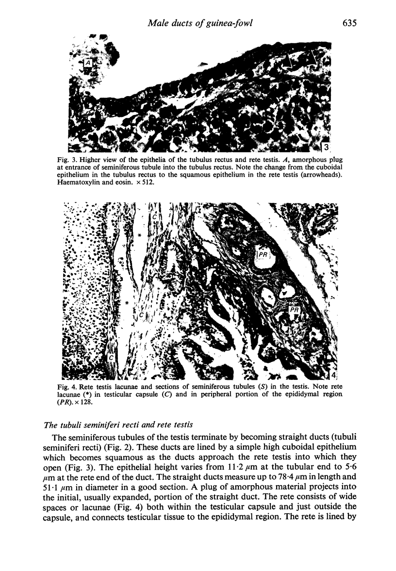
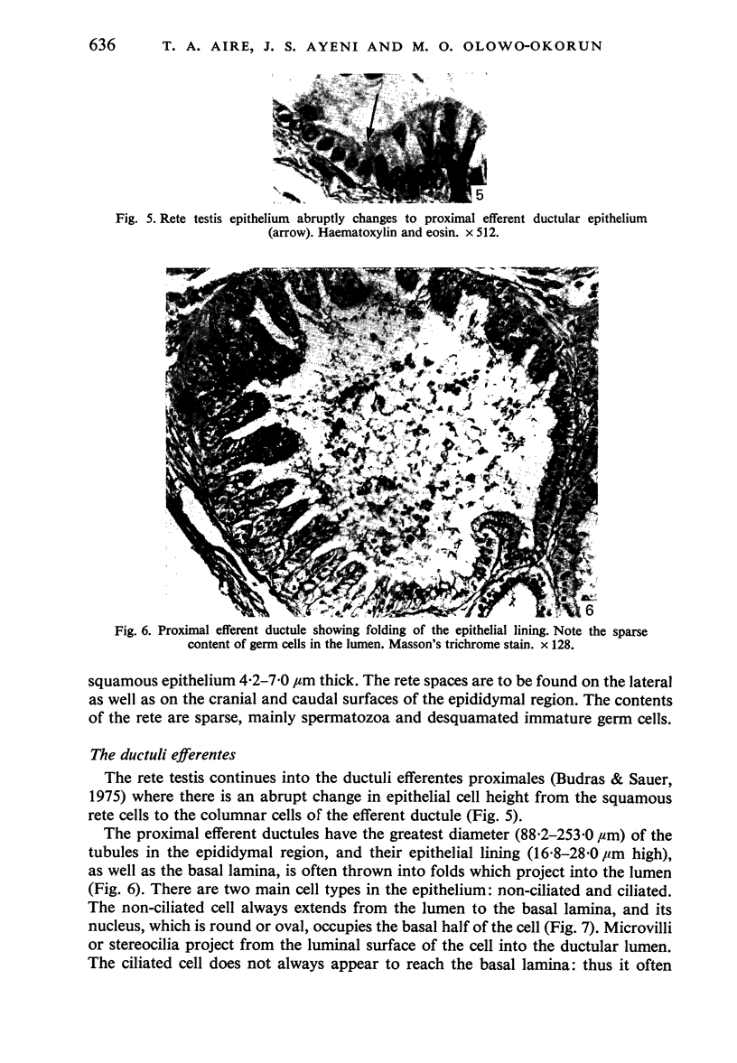
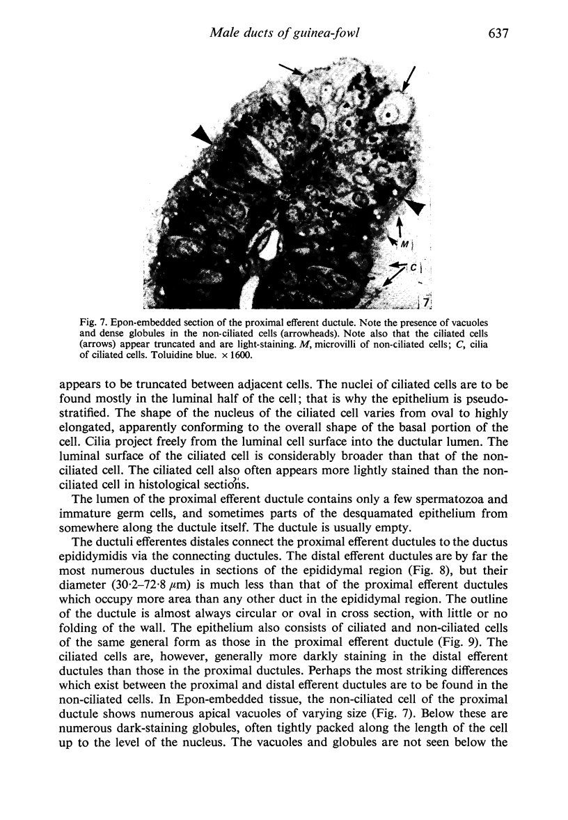
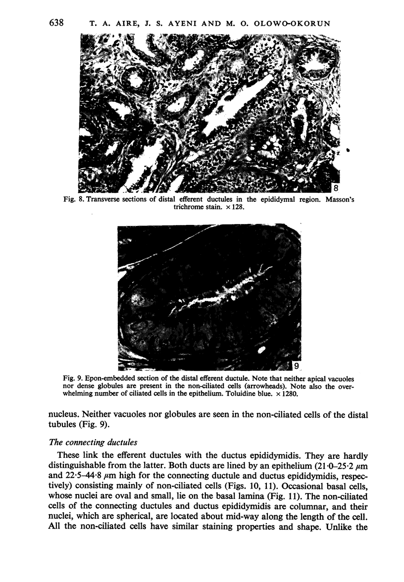
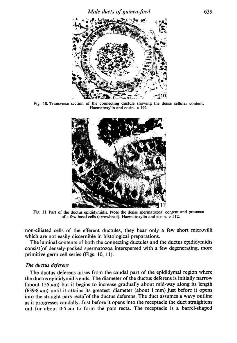
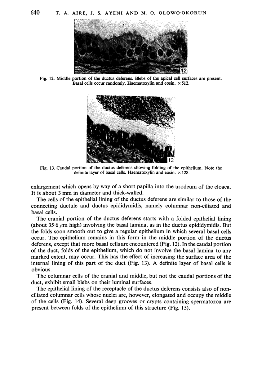
Images in this article
Selected References
These references are in PubMed. This may not be the complete list of references from this article.
- Aire T. A. The epididymal region of the Japanese quail (Coturnix coturnix japonica). Acta Anat (Basel) 1979;103(3):305–312. [PubMed] [Google Scholar]
- Budras K. D., Sauer T. Morphology of the epididymis of the cock (Gallus domesticus) and its effect upon the steroid sex hormone synthesis. I. Ontogenesis, morphology and distribution of the epididymis. Anat Embryol (Berl) 1975 Dec 23;148(2):175–196. doi: 10.1007/BF00315268. [DOI] [PubMed] [Google Scholar]
- Hess R. A., Thurston R. J., Biellier H. V. Morphology of the epididymal region and ductus deferens of the turkey (Meleagris gallopavo). J Anat. 1976 Nov;122(Pt 2):241–252. [PMC free article] [PubMed] [Google Scholar]
- Hess R. A., Thurston R. J. Ultrastructure of epithelial cells in the epididymal region of the turkey (Meleagris gallopavo). J Anat. 1977 Dec;124(Pt 3):765–778. [PMC free article] [PubMed] [Google Scholar]
- LADMAN A. J., YOUNG W. C. An electron microscopic study of the ductuli efferentes and rete testis of the guinea pig. J Biophys Biochem Cytol. 1958 Mar 25;4(2):219–226. doi: 10.1083/jcb.4.2.219. [DOI] [PMC free article] [PubMed] [Google Scholar]
- LAKE P. E. The male reproductive tract of the fowl. J Anat. 1957 Jan;91(1):116–129. [PMC free article] [PubMed] [Google Scholar]
- STOLL R., MARAUD R. Sur la constitution de l'épididyme du coq. C R Seances Soc Biol Fil. 1955 Apr;149(7-8):687–689. [PubMed] [Google Scholar]
- Tingari M. D. On the structure of the epididymal region and ductus deferens of the domestic fowl (Gallus domesticus). J Anat. 1971 Sep;109(Pt 3):423–435. [PMC free article] [PubMed] [Google Scholar]
- Tingari M. D. The fine structure of the epithelial lining of the ex-current duct system of the testis of the domestic fowl (Gallus domesticus). Q J Exp Physiol Cogn Med Sci. 1972 Jul;57(3):271–295. doi: 10.1113/expphysiol.1972.sp002162. [DOI] [PubMed] [Google Scholar]


