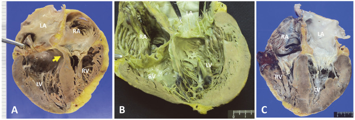
Figure 3.
Representative longitudinal section of the right and left atriums and ventricles (4-chamber view). (A) The summit of the ventricular septum is regionally thinned with fibrosis. The thinned wall curves to the right ventricle, which is affected by blood flow pressure (yellow arrow; case no. 8, 86-year-old woman; posterior-anterior side view). The anterior wall of the atrioventricular junction beneath the mitral valve is consecutively thinned, extending from the thinned septal lesion. (B) The ventricular septal wall is completely thinned from the atrioventricular junction to the apex edge (case no. 3, 64-year-old man; anterior-posterior side view). Thinned lesions extend to both ventricles. Black colored lesions indicate radiofrequency ablation sites for eliminating ventricular tachycardia. (C) The thickness of the ventricular septal wall is well preserved (case no. 2, 63-year-old man). LA, left atrium; LV, left ventricle; RA, right atrium; RV, right ventricle.

