Abstract
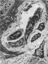
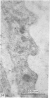
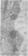
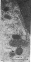
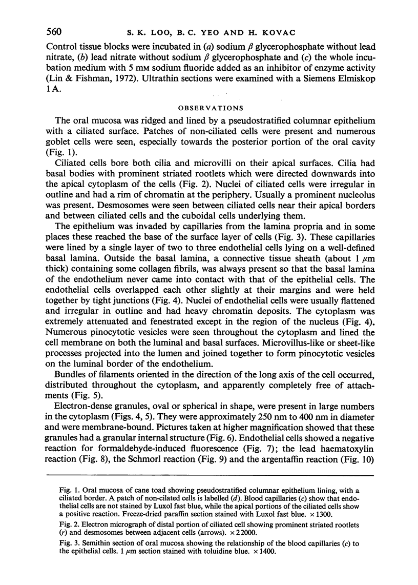
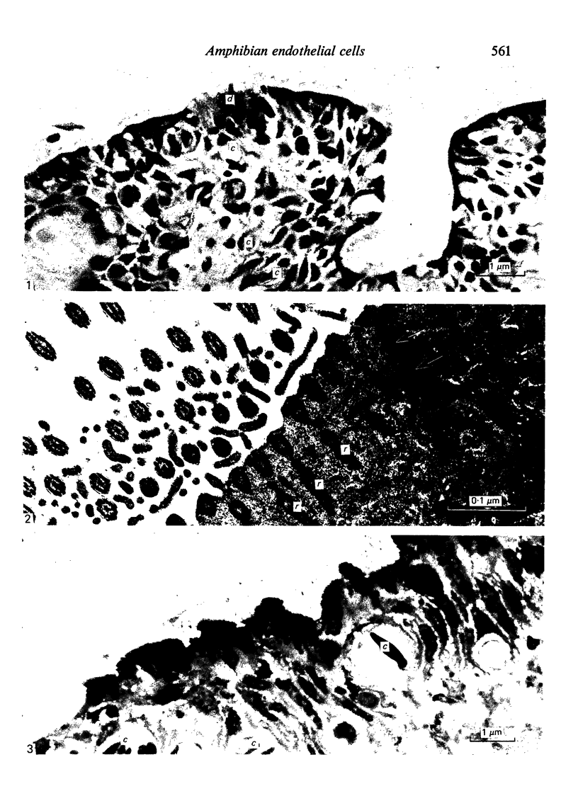
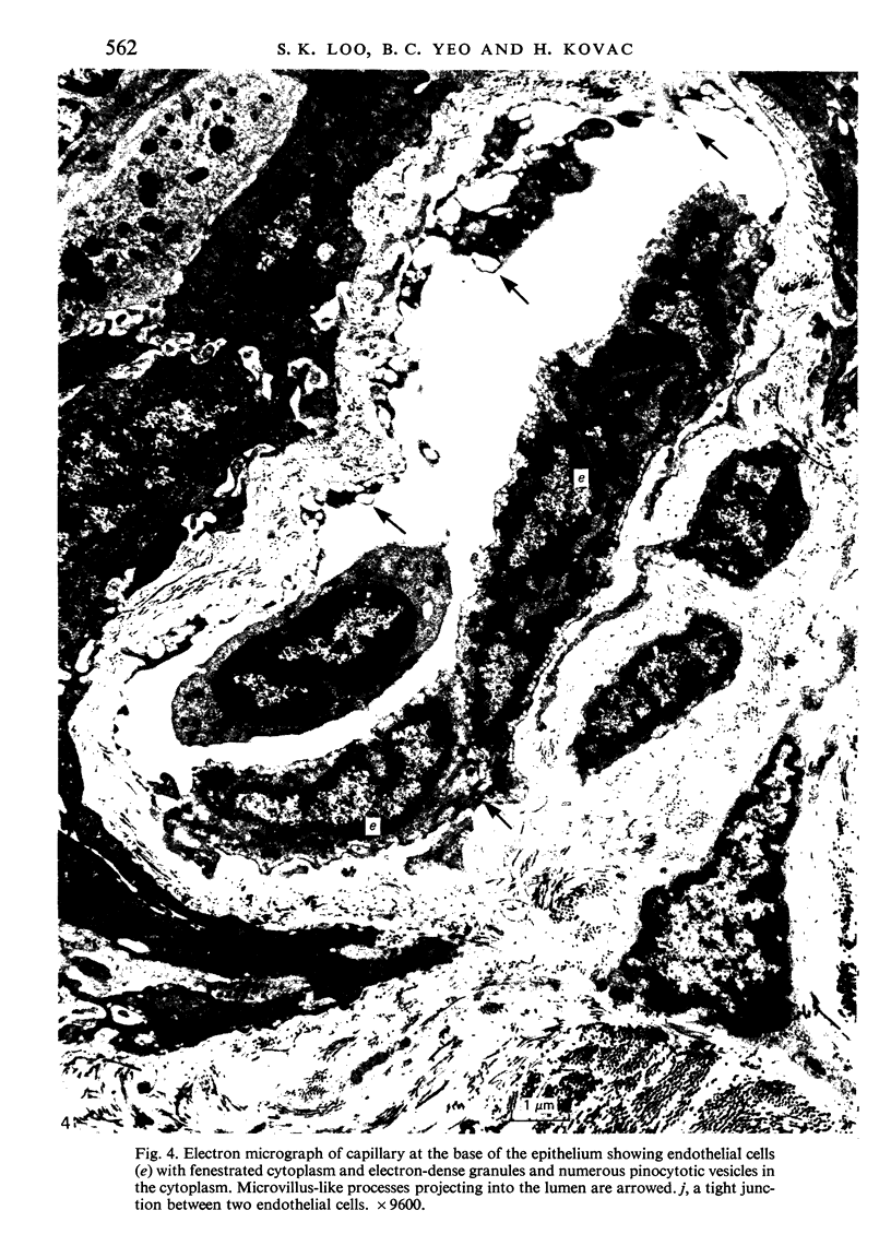
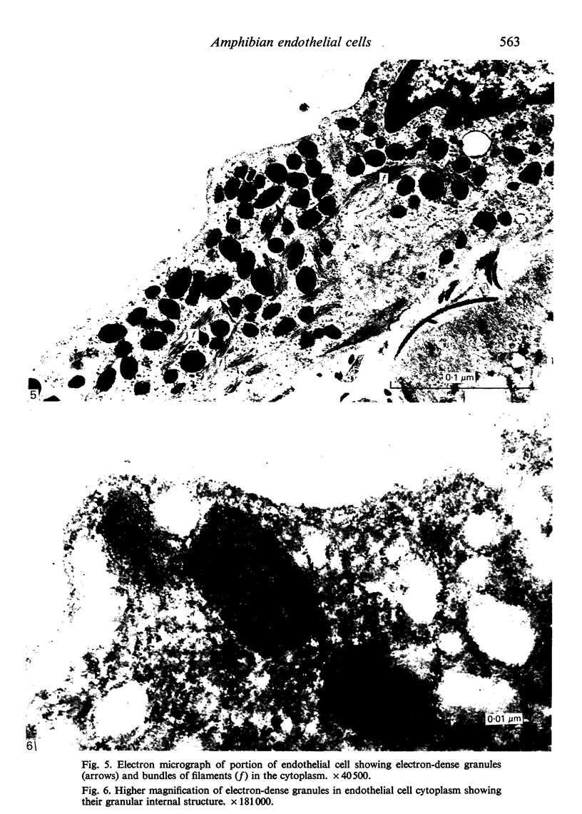
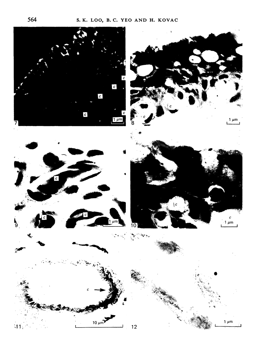
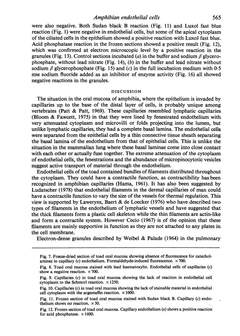
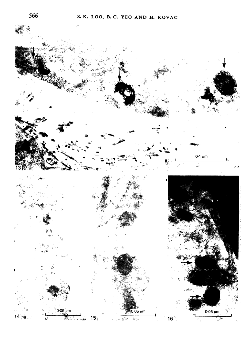
The oral mucosa of the cane toad (Bufo marinus) is lined by a pseudostratified columnar ciliated epithelium containing an intraepithelial network of capillaries, which penetrates it to the bases of the distal layer of cells. The capillaries are lined by fenestrated endothelium lying on a complete basal lamina. A connective tissue sheath, approximately 1 micrometer thick, surrounds the capillaries and separates them from the surrounding epithelial cells. Endothelial cells resemble those in lymphatic capillaries in that they show microvillus-like processes or folds projecting into the lumen and also have extremely attenuated and fenestrated cytoplasm except in the nuclear region. Numerous pinocytotic vesicles, bundles of filaments and many electrondense granules occur in the cytoplasm. These granules are oval or round in shape and approximately 250-400 micrometer in diameter. Histochemical tests on the endothelial cells show that the granules do not contain pigment, as both the Schmorl and argentaffin reactions are negative. Both the Sudan black B and Luxol fast blue reactions are also negative showing the lack of stainable lipids. The formaldehyde-induced fluorescence, the argentaffin reactions and lead haematoxylin reactions are negative, indicating that they do not have the characteristics of endocrine cells. The acid phosphatase reaction gives a positive result, localized to the site of the granules by electron microscopy and suggesting that these granules in amphibian capillaries may have a lysosomal function.
Full text
PDF
Images in this article
Selected References
These references are in PubMed. This may not be the complete list of references from this article.
- Bertini F., Piezzi R., Gutierrez L. Further studies on endothelial cells of vertebrates and the problem of endothelial granules. Experientia. 1972 Nov 15;28(11):1350–1352. doi: 10.1007/BF01965339. [DOI] [PubMed] [Google Scholar]
- Bertini F., Santolaya R. A novel type of granules observed in toad endothelial cells and their relationship with blood pressure active factors. Experientia. 1970 May 15;26(5):522–523. doi: 10.1007/BF01898486. [DOI] [PubMed] [Google Scholar]
- Cecio A. Ultrastructural features of cytofilaments within mammalian endothelial cells. Z Zellforsch Mikrosk Anat. 1967;83(1):40–48. doi: 10.1007/BF00334737. [DOI] [PubMed] [Google Scholar]
- Gorgas K., Böck P., Tischendorf F., Curri S. B. The fine structure of human digital arterio-venous anastomoses (Hoyer-Grosser's organs). Anat Embryol (Berl) 1977 May 12;150(3):269–289. doi: 10.1007/BF00318346. [DOI] [PubMed] [Google Scholar]
- HAMA K. On the existence of filamentous structures in endothelial cells of the amphibian capillary. Anat Rec. 1961 Mar;139:437–441. doi: 10.1002/ar.1091390313. [DOI] [PubMed] [Google Scholar]
- Hruban Z., Vigil E. L., Slesers A., Hopkins E. Microbodies: constituent organelles of animal cells. Lab Invest. 1972 Aug;27(2):184–191. [PubMed] [Google Scholar]
- Iijima T., Wasano T. Occurrence of catecholamine-containing specific granules in the venous endothelia of carp. Am J Anat. 1978 Sep;153(1):171–176. doi: 10.1002/aja.1001530112. [DOI] [PubMed] [Google Scholar]
- Lauweryns J. M., Baert J., De Loecker W. Fine filaments in lymphatic endothelial cells. J Cell Biol. 1976 Jan;68(1):163–167. doi: 10.1083/jcb.68.1.163. [DOI] [PMC free article] [PubMed] [Google Scholar]
- Lemeunier A., Burri P. H., Weibel E. R. Absence of acid phosphatase activity in specific endothelial organelles. Histochemie. 1969;20(2):143–149. doi: 10.1007/BF00268708. [DOI] [PubMed] [Google Scholar]
- Lin C. W., Fishman W. H. Microsomal and lysosomal acid phosphatase isoenzymes of mouse kidney. Characterization and separation. J Histochem Cytochem. 1972 Jul;20(7):487–498. doi: 10.1177/20.7.487. [DOI] [PubMed] [Google Scholar]
- Ludatscher R. M. Ultrastructure of human dermal blood vessels with special reference to the endothelial filaments. Virchows Arch B Cell Pathol. 1978 Jun 19;27(4):347–357. doi: 10.1007/BF02889006. [DOI] [PubMed] [Google Scholar]
- Pearse A. G. The diffuse neuroendocrine system and the apud concept: related "endocrine" peptides in brain, intestine, pituitary, placenta, and anuran cutaneous glands. Med Biol. 1977 Jun;55(3):115–125. [PubMed] [Google Scholar]
- Piezzi R. S., Santolaya R. C., Bertini F. The fine structure of endothelial cells of toad arteries. Anat Rec. 1969 Oct;165(2):229–231. doi: 10.1002/ar.1091650208. [DOI] [PubMed] [Google Scholar]
- SEVIER A. C., MUNGER B. L. TECHNICAL NOTE: A SILVER METHOD FOR PARAFFIN SECTIONS OF NEURAL TISSUE. J Neuropathol Exp Neurol. 1965 Jan;24:130–135. doi: 10.1097/00005072-196501000-00012. [DOI] [PubMed] [Google Scholar]
- Santolaya R. C., Bertini F. Fine structure of endothelial cells of vertebrates. Distribution of dense granules. Z Anat Entwicklungsgesch. 1970;131(2):148–155. doi: 10.1007/BF00523293. [DOI] [PubMed] [Google Scholar]
- Solcia E., Capella C., Vassallo G. Lead-haematoxylin as a stain for endocrine cells. Significance of staining and comparison with other selective methods. Histochemie. 1969;20(2):116–126. doi: 10.1007/BF00268705. [DOI] [PubMed] [Google Scholar]
- Spurr A. R. A low-viscosity epoxy resin embedding medium for electron microscopy. J Ultrastruct Res. 1969 Jan;26(1):31–43. doi: 10.1016/s0022-5320(69)90033-1. [DOI] [PubMed] [Google Scholar]
- Stehbens W. E. Ultrastructure of vascular endothelium in the frog. Q J Exp Physiol Cogn Med Sci. 1965 Oct;50(4):375–384. doi: 10.1113/expphysiol.1965.sp001804. [DOI] [PubMed] [Google Scholar]
- Tabuchi H., Yamamoto T. Specific granules in the endothelia of blood and lymphatic vessels in the cardiac valves of dogs. Arch Histol Jpn. 1974 Nov;37(3):217–224. doi: 10.1679/aohc1950.37.217. [DOI] [PubMed] [Google Scholar]
- WEIBEL E. R., PALADE G. E. NEW CYTOPLASMIC COMPONENTS IN ARTERIAL ENDOTHELIA. J Cell Biol. 1964 Oct;23:101–112. doi: 10.1083/jcb.23.1.101. [DOI] [PMC free article] [PubMed] [Google Scholar]


