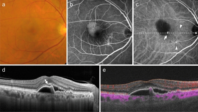
Fig. 4.
Polypoidal choroidal vasculopathy. a Fundus photo shows round PED. b FA shows dye pooling in the area of PED. Scale bar, 200 μm. c ICGA shows PED as a fluorescence block, with type 1 MNV (arrowhead) and polypoidal lesions (arrow). Scale bar, 200 μm. d OCT corresponding to the site indicated by the dashed arrow in c shows a serous PED with a notch (arrow) and a shallow elevation of the RPE with a double layer sign. e B-scan OCTA image shows blood flow signals (pink) at the site of the notch

