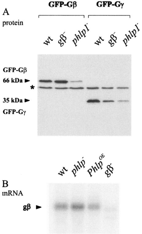
FIG. 2.
Expression of Gβ and Gγ in phlp1− cells. (A) Steady-state protein levels of GFP-Gβ and GFP-Gγ are reduced in phlp1− cells. Wild-type AX3 cells (wt) and gβ− and phlp1− knockout cells were transfected with plasmids encoding GFP-Gβ or GFP-Gγ. Cell lysates were prepared and analyzed through Western blot analysis with a GFP antibody. The molecular masses of GFP-Gβ and GFP-Gγ are indicated. The asterisk denotes a cross-reacting AX3 band. (B) Northern blots show normal expression of Gβ in phlp1− cells. Poly(A) mRNA was isolated from wild-type AX3 cells (wt), phlp1− knockout cells, phlp1− cells overexpressing PhLP1 (PhlpOE), and gβ− knockout cells. RNA was size fractionated, transferred, and probed with gβ.

