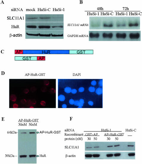
FIG. 4.
SLC11A1 expression is inhibited and can be restored in HuR knockdown cells. (A) HL-60 cells were transiently transfected with siRNA targets HuR (HuSi-1), control siRNA (HuSi-C, a mutant of HuSi-1), or mock (transfection reagents only). Four h after transfection, HL-60 cells were treated with PMA (10 ng/ml) for 68 h. Protein extracts of mock-transfected HL-60 cells, as well as those of HuSi-C- and HuSi-1-transfected HL-60 cells, were prepared and analyzed by Western blotting with antibodies specific to SLC11A1, HuR, and β-actin. (B) HL-60 cells were transfected with siRNA and then treated with PMA as described above. Forty-eight and 72 h after PMA treatment, total RNA from HL-60 cells transfected with HuSi-1, HuSi-C, or mock were isolated, and Northern blot analysis was performed to determine SLC11A1 mRNA levels. (C) Schematic representations of fusion proteins AP-HuR-GST and GST-HuR (a control) used to rescue HuR siRNA-treated HL-60 cells. (D) The Alexa Fluor 594-labeled AP-HuR-GST was added to HL-60 cells 4 h after transfection with HuSi-1. Six h after the addition of labeled AP-HuR-GST, the cells were washed, fixed (4% paraformaldehyde; 15 min), and visualized with a Zeiss Axiovision 3.1 microscope. (E) HL-60 cells were transfected with HuSi-1. The recombinant protein AP-HuR-GST was added to cells 4 h posttransfection, and total protein extracts were prepared 6 h after the addition of AP-HuR-GST recombinant protein. Western blot analysis was performed to detect the absorbance of AP-HuR-GST by HL-60 cells. (F) HL-60 cells were transfected with HuSi-1 or HuSi-C, and 4 h after transfection, recombinant proteins GST-AP and AP-HuR-GST were added to the cells. Six h later, cells were treated with PMA (10 ng/ml). Seventy-two h after HuR siRNA treatment, total protein extracts were prepared. Western blot analysis was performed using antibodies specific to SLC11A1 and β-actin.

