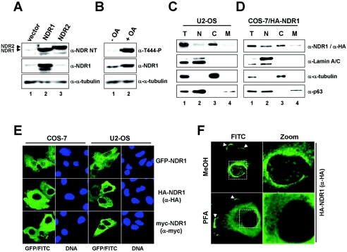
FIG. 1.
Human NDR1 is a mainly cytoplasmic kinase. A. U2-OS cells expressing empty vector (lane 1), untagged human NDR1 (lane 2), or untagged human NDR2 (lane 3) were processed for immunoblotting using anti-NDR NT (top panel), anti-NDR1 (middle panel), and anti-α-tubulin (bottom panel) antibodies. Arrowheads indicate the positions of NDR1 and NDR2. B. U2-OS cells were incubated in the absence (lane 1) or presence (lane 2) of OA before processing for Western blotting using anti-Thr444-P (top panel), anti-NDR1 (middle panel), and anti-α-tubulin (bottom panel) antibodies. C. U2-OS cells were subjected to biochemical cell fractionation (T, total; N, nuclear; C, cytoplasmic; M, membrane) and processed for immunoblotting with anti-NDR1 (top panel), anti-lamin A/C (nuclear marker; top middle panel), anti-α-tubulin (cytoplasmic marker; bottom middle panel), and anti-p63/Climp (perinuclear/membrane marker; bottom panel) antibodies. D. COS-7 cells expressing HA-tagged NDR1 were subjected to biochemical cell fractionation (T, total; N, nuclear; C, cytoplasmic; M, membrane) and processed for immunoblotting with anti-HA (top panel), anti-lamin A/C (top middle panel), anti-α-tubulin (bottom middle panel), and anti-p63/Climp (bottom panel) antibodies. E. COS-7 and U2-OS cells expressing either GFP-NDR1 (top panels), HA-NDR1 (middle panels), or myc-NDR1 (bottom panels) were analyzed by indirect immunofluorescence microscopy using anti-HA Y11 (middle panels), anti-myc (bottom panels), or no (top panels) antibody. GFP/FITC signals are shown in green. Nuclei are in blue. F. COS-7 cells expressing HA-NDR1 were fixed with either methanol (MeOH) (top panels) or paraformaldehyde (PFA) (bottom panels) before permeabilization and antibody staining using anti-HA Y11 antibody. Arrowheads indicate NDR1 species found in plasma membrane structures. Enlargements of the regions indicated by white rectangles are shown on the right (Zoom).

