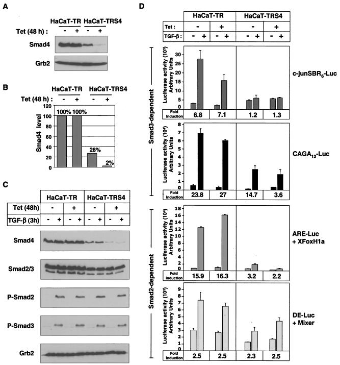
FIG. 1.
Characterization of a stable HaCaT cell line expressing a Tet-inducible siRNA targeted against Smad4. (A and B) Knockdown of Smad4 is strongly induced by Tet in HaCaT-TRS4 cells. HaCaT-TR parental cells and HaCaT-TRS4 cells were treated with Tet for 48 h, and equal amounts of protein were analyzed by Western blotting using antibodies against Smad4 and Grb2 as a loading control (A). Smad4 levels were quantified using the Odyssey detection system (B). (C) Knockdown of Smad4 has no effect on Smad2/3 phosphorylation after TGF-β stimulation. HaCaT-TR parental cells and HaCaT-TRS4 cells were treated with Tet for 48 h and then with TGF-β for 3 h. Equal amounts of protein were analyzed by Western blotting using antibodies against Smad4, Smad2/3, phosphorylated Smad2 (P-Smad2), phosphorylated Smad3 (P-Smad3), and Grb2 as a loading control. (D) Knockdown of Smad4 has differential effects on TGF-β-induced transcription from Smad3-dependent and Smad2-dependent synthetic reporters. Cells were transfected with the Smad3-dependent reporters CAGA12-Luc or c-junSBR6-Luc and the Smad2-dependent reporters ARE-Luc and a plasmid expressing XFoxH1a/XFast-1 or DE-Luc and a plasmid expressing Mixer. Where appropriate, Tet was added at the time of the transfection. Cells were then incubated for 40 h, and TGF-β was added 8 h before assaying luciferase activity. The TGF-β fold inductions are given at the bottom of each graph.

