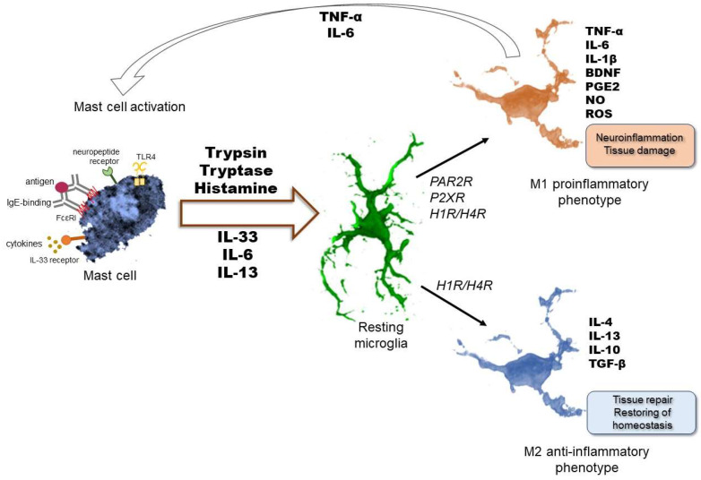
Figure 1.
Interactions between mast cells and microglia in the brain. Bidirectional communication may influence not only the release of different mediators but also impact the state of activation of these immune cells. Microglial phenotype switching is influenced by signals arising from their environment. Activated mast cells could be a source of these mediators. Mast cell-derived factors, like different interleukins, trypsin, tryptase, or histamine, trigger the alteration of the microglial phenotype to switch either to M1 pro-inflammatory or M2 anti-inflammatory subclasses. Both the M1 and M2 phenotypes can be characterized by a specific spectrum of pro- or anti-inflammatory mediator release leading to neuroinflammation or tissue repair, respectively. Activated microglia may then affect mast cell activation, resulting in a more prominent inflammatory response. FcεR1: high-affinity immunoglobulin E (IgE) receptor, TLR4: toll-like receptor 4, IL: interleukin, PAR2R: protease-activated receptor 2, P2XR: purinergic P2 receptor, H1R/H4R: histamine receptor 1 and 4, TNF-α: tumor necrosis factor-α, TGF-β: transforming growth factor-β, BDNF: brain-derived neurotrophic factor, PGE2: prostaglandin E2, NO: nitric oxide, ROS: reactive oxygen species.

