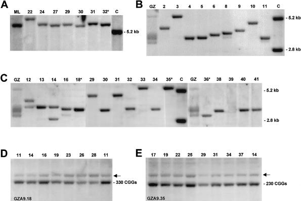
Figure 1.
Southern analysis of fragile X expansions harbored in murine A9 host cells. DNA samples isolated from fibroblasts of donors (ML and GZ) and from A9 hybrid clones were cleaved by restriction enzyme and hybridized to probe Ox1.9. A, Discrete-length alleles of methylated full expansions were isolated into different hybrid clones. DNA samples were cleaved with EcoRI plus EagI. The results for the donor’s (ML) fibroblasts and for six hybrid clones are shown with the clone numbers indicated. Because of methylation of the EagI site in the FMR1 promoter, the expanded fragments are larger than the normal 5.2-kb fragment seen in the control (lane C). Clone 32* was selected for microcell generation. B and C, Unmethylated expansions isolated into A9 hybrid clones (numbers shown above the lanes) and visualized by hybridization to EcoRI-plus-EagI–cleaved DNA. Cleavage of unmethylated EagI sites of expanded alleles results in fragments between 2.8 and 5.2 kb. Unmethylated 2.8-kb and methylated 5.2-kb-fragments are carried on the active and inactive X chromosomes of the female control (lane C). Whereas the donor’s (GZ) fibroblasts presented smears of multiple expansions, the majority of hybrids harbored a single expanded fragment with unmethylated EagI site. Clones 18*, 35*, and 36* were used in further experiments. D and E, Mitotic stability of expansions isolated from GZ into A9 clones 18 and 35 (GZA9.18, GZA9.35). Clonal DNAs were isolated at successive passages and cleaved with HindIII. The passage numbers are given above the lanes in D and E; population doublings are three to four times the passage number. The HindIII fragments of the murine gene, indicated by arrows (←), are seen in D and E above the human fragments carrying 330 and 230 CGGs, respectively.

