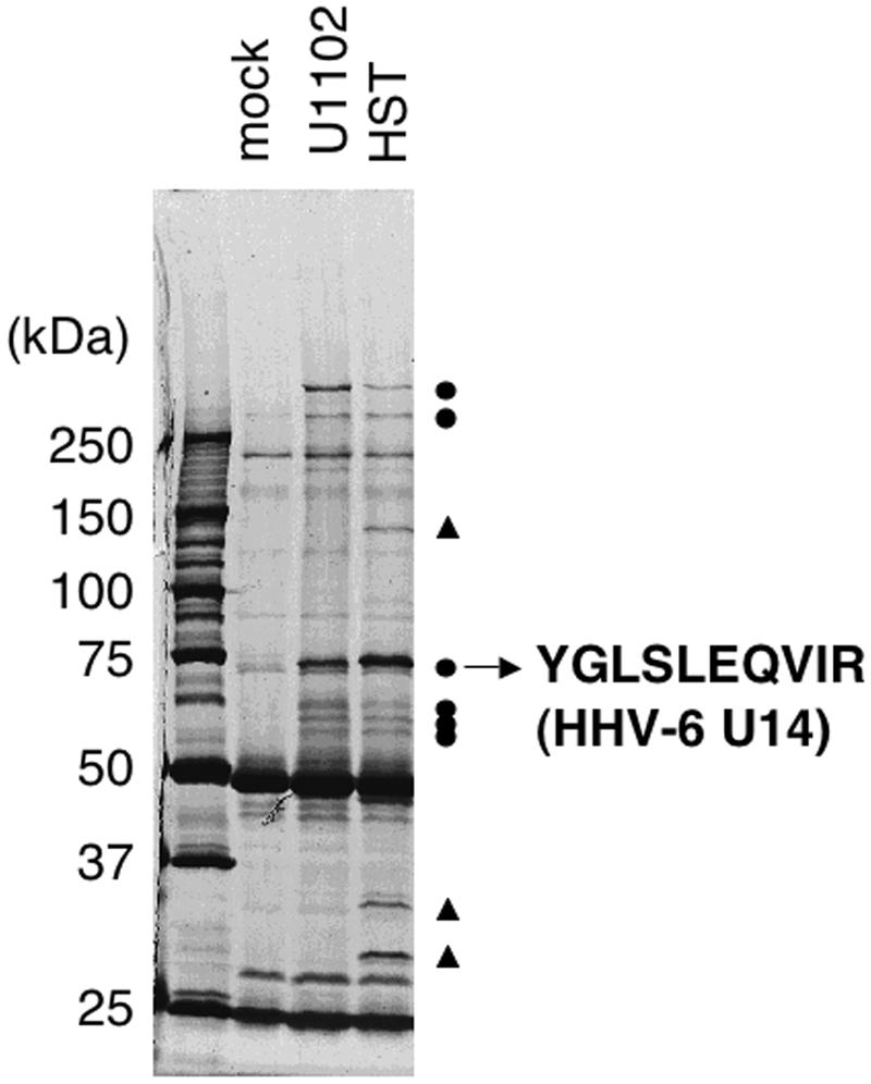
FIG. 1.

Coimmunoprecipitation experiments with the anti-p53 MAb and identification of the 75-kDa protein as HHV-6 U14. Molt-3 cells infected with the U1102 or HST strain were lysed with TNE buffer at 24 hpi and subjected to coimmunoprecipitation. Samples were resolved on 6% SDS-PAGE, the proteins were stained with Coomassie brilliant blue, and the protein band of interest was excised from the gel (the photo is of a silver-stained gel). Circles and triangles indicate six bands common to both variants and three bands specific to HHV-6B HST, respectively.
