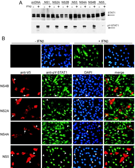
FIG. 5.
Identification of LGTV nonstructural proteins that function as IFN antagonists. (A) Vero cells expressing LGTV NS proteins fused to V5 epitope tags were treated with blasticidin to obtain a cell population, the majority of which expressed the individual NS protein indicated, or the backbone plasmid, pcDNA6.2/V5. Cells were then treated with IFN-β and examined for STAT1 phosphorylation by Western blot analysis. Steady-state STAT1 levels (upper panel) and tyrosine-phosphorylated STAT1 (pY-STAT1) (lower panel) are shown in cell populations expressing LGTV NS proteins and either left untreated (−) or treated (+) with IFN-β. (B) Vero cells were treated with IFN-β, fixed in methanol, and stained for anti-V5 (red) and anti-pY-STAT1 (green). Nuclei were counterstained with DAPI (blue). The upper panel demonstrates the localization of tyrosine-phosphorylated STAT1 in the nuclei of untransfected cells that were left untreated or treated with IFN-β. The lower panels represent cells that express NS4B, NS2A, NS4A, and NS5. Examples of V5-positive and either pY-STAT1-positive (arrows) or pY-STAT1-negative (arrowheads) cells are indicated.

