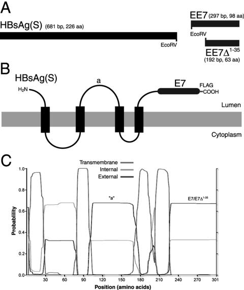
FIG. 1.
Schematic representation of HBsAg(S)16EE7 fusion genes and proteins and their expression in mammalian cells. (A) HBsAg(S)/E7 fusion genes used in this study. The stop codon of HBsAg(S) was replaced by an EcoRV site, which allows for in-frame 3′ end fusion of coding sequences. Constructs were derived by fusion of HBsAg(S) to either a complete EE7 gene or a truncated mutant devoid of the first 35 codons (EE7Δ1-35). A FLAG tag (not represented) was added at the 3′ end of the EE7 sequences. (B) Topology of the HBsAg(S)-HPV-16E7 fusion proteins in the ER membrane. The luminal side of the protein is that exposed on the surface of the extruded particles. (C) Transmembrane domain prediction of the HBsAg-E7 fusion proteins according to the algorithm developed by Sonnhammer et al. (37). The positions of the “a” determinant and the E7 domains are indicated.

