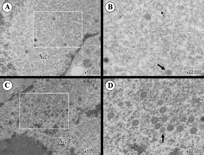
FIG. 4.
VZV virion formation in melanoma cells in vitro. (A) Virions were clearly distinguishable in the nucleus of pOka-infected melanoma cells. (B) Magnification of pOka virions from the boxed area of panel A shows the presence of intranuclear virions (large arrows) and empty capsids (small arrows). (C) Virions were also abundantly present in the nucleus of pOka66S-infected melanoma cells. (D) Magnification of the boxed area of panel C shows that these nucleocapsids have a normal appearance comparable to those in pOka-infected cells. The magnification and locations of cytoplasm (cyt) and nucleus (nuc) are indicated.

